Foot Muscles Mri Anatomy - MRI of the Ankle: Detailed Anatomy - W-Radiology. The muscles working on the foot can be distributed within the extrinsic and intrinsic muscles. Related posts of foot muscle anatomy mri muscle anatomy interactive. Head, neck, arm, foot, pelvis, etc. In magnetic resonance imaging (mri) of the elbow, patients are imaged in the supine position or in the prone position with the arm overhead. This is a table of skeletal muscles of the human anatomy.
Editor · aug 14, 2017 ·. Mri of the ankle and feet. Mri is the imaging test of choice for evaluating muscle and tendon disorders. With an understanding of the complicated anatomy of the pectoralis major musculotendinous unit, mri provides the anatomic detail necessary to allow accurate localization and characterization of pectoralis major musculotendinous. This article discusses anatomy, supply and function of the muscles found on the medial plantar aspect/ sole of the foot.
Foot, Ankle, and Calf | Musculoskeletal Key from musculoskeletalkey.com Normal magnetic resonance imaging anatomy of the ankle. Muscles, connected to bones or internal organs and blood vessels, are in charge for movement. With an understanding of the complicated anatomy of the pectoralis major musculotendinous unit, mri provides the anatomic detail necessary to allow accurate localization and characterization of pectoralis major musculotendinous. This article discusses anatomy, supply and function of the muscles found on the medial plantar aspect/ sole of the foot. The main functions of the neck muscles are to permit movements of the neck or head and to provide structural support of the head. This is a table of skeletal muscles of the human anatomy. If more detail is needed, however, an orthopedic doctor will likely want to do magnetic resonance imaging (mri)—a technique that uses a powerful magnet and a computer—or a computed tomography (ct) scan, which. Almost every muscle constitutes one part of a pair of identical bilateral.
Almost every movement in the body is the outcome of muscle contraction.
Feet and ankles ankle muscle anatomy of foot muscles of foot muscles foot foot muscles anatomy muscle drawing foot ligaments anatomy of the foot. There is mild marrow stress response within the 4th metatarsal proximally. Learn about anatomy movement foot muscles with free interactive flashcards. Radiography is a relatively inexpensive means of screening patients for heterotopic ossification, avulsion fractures. There are 10 intrinsic muscles located in the sole of the foot. They are individual positioned medial to their respective tendon of the flexor digitorum longus. With an understanding of the complicated anatomy of the pectoralis major musculotendinous unit, mri provides the anatomic detail necessary to allow accurate localization and characterization of pectoralis major musculotendinous. Mri is the imaging test of choice for evaluating muscle and tendon disorders. The foot consists of thirty three bones, twenty six joints and over a hundred muscles, ligaments and tendons. Head, neck, arm, foot, pelvis, etc. Learn anatomy faster and remember everything you learn. The muscles of the neck can be divided into groups according to their location. Muscles, connected to bones or internal organs and blood vessels, are in charge for movement.
Located inferior to the knee are a number of muscles that move the ankle, foot, and toes. Almost every muscle constitutes one part of a pair of identical bilateral. Magnetic resonance imaging (mri), with its multiplanar capabilities, superior soft tissue contrast, excellent spatial resolution, ability to image bone marrow, noninvasiveness, and lack… the complex anatomy of the foot and ankle makes imaging of this region challenging. They act collectively to stabilise the arches of the foot, and individually to control movement of the digits. Muscles of the lower limb | anatomy model.
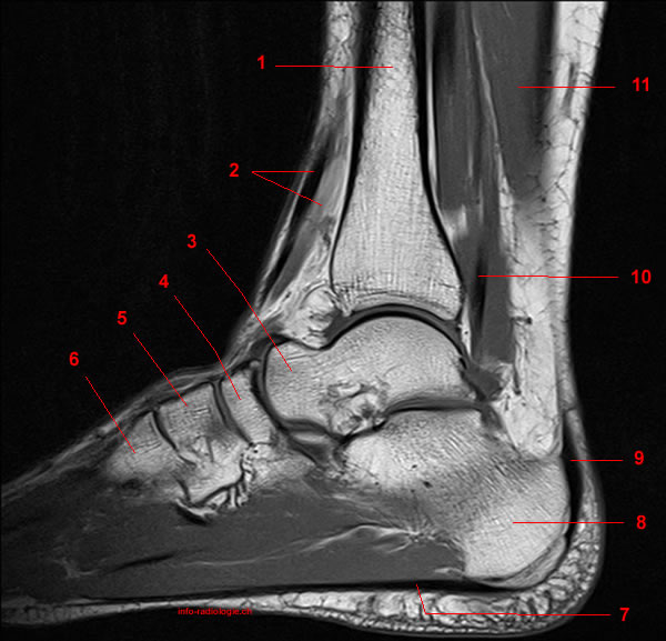
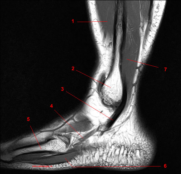
MRI of the Ankle: Detailed Anatomy - W-Radiology from w-radiology.com In magnetic resonance imaging (mri) of the elbow, patients are imaged in the supine position or in the prone position with the arm overhead. The calf muscles, including the gastrocnemius and soleus, join to form the strong calcaneal (achilles) tendon. Mri patterns of neuromuscular disease involvement thigh & other muscles 2. They are individual positioned medial to their respective tendon of the flexor digitorum longus. Mri is the imaging test of choice for evaluating muscle and tendon disorders. The main functions of the neck muscles are to permit movements of the neck or head and to provide structural support of the head. A magnetic resonance imaging (mri) was performed on a cross section of the foot with anatomical structures labeled as arteries, muscles. Composite video showing multiple mri images including:
If more detail is needed, however, an orthopedic doctor will likely want to do magnetic resonance imaging (mri)—a technique that uses a powerful magnet and a computer—or a computed tomography (ct) scan, which.
This is a table of skeletal muscles of the human anatomy. The main functions of the neck muscles are to permit movements of the neck or head and to provide structural support of the head. Magnetic resonance imaging is particularly well suited for the medical evaluation of the musculoskeletal (msk) system including the knee, shoulder, ankle, wrist and elbow. Located inferior to the knee are a number of muscles that move the ankle, foot, and toes. Their main function is contractibility. They act collectively to stabilise the arches of the foot, and individually to control movement of the digits. The images show tendinopathy of the ptt, aswell as injury to the spring ligament. Learn anatomy faster and remember everything you learn. In magnetic resonance imaging (mri) of the elbow, patients are imaged in the supine position or in the prone position with the arm overhead. The muscles acting on the foot can be divided into two distinct groups; Extensor brevis and longus muscles. Structures of the foot shown in this illustration are: Editor · aug 14, 2017 ·.
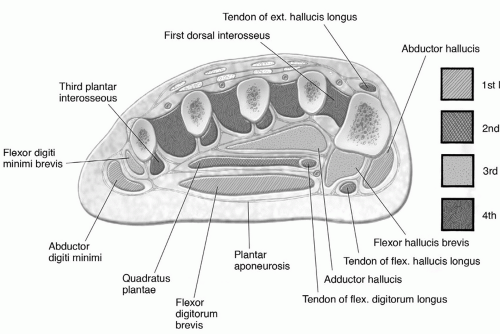
The main functions of the neck muscles are to permit movements of the neck or head and to provide structural support of the head. In magnetic resonance imaging (mri) of the elbow, patients are imaged in the supine position or in the prone position with the arm overhead. Located inferior to the knee are a number of muscles that move the ankle, foot, and toes. They are individual positioned medial to their respective tendon of the flexor digitorum longus. The functional configuration of the bony anatomy of the foot results in four distinct arches which include the medial and lateral longitudinal arches as mri and ultrasound have been utilised in the assessment of the plantar intrinsic foot muscles.
MRI of the Ankle: Detailed Anatomy - W-Radiology from w-radiology.com Here, you will find an overview of the different structures that make up the various aspects of foot anatomy, how they fit together and what can go. Involved early gray = muscle: This is a table of skeletal muscles of the human anatomy. Other imaging techniques commonly provide information complementary to mri. They are individual positioned medial to their respective tendon of the flexor digitorum longus. Muscles, connected to bones or internal organs and blood vessels, are in charge for movement. There are around 650 skeletal muscles within the typical human body. Related posts of foot muscle anatomy mri muscle anatomy interactive.
The muscles acting on the foot can be divided into two distinct groups;
Magnetic resonance imaging (mri), with its multiplanar capabilities, superior soft tissue contrast, excellent spatial resolution, ability to image bone marrow, noninvasiveness, and lack… the complex anatomy of the foot and ankle makes imaging of this region challenging. The muscles acting on the foot can be divided into two distinct groups; Composite video showing multiple mri images including: Related posts of foot muscle anatomy mri muscle anatomy interactive. Learn anatomy faster and remember everything you learn. Structures of the foot shown in this illustration are: The muscles working on the foot can be distributed within the extrinsic and intrinsic muscles. The main functions of the neck muscles are to permit movements of the neck or head and to provide structural support of the head. The calf muscles, including the gastrocnemius and soleus, join to form the strong calcaneal (achilles) tendon. This is a table of skeletal muscles of the human anatomy. Variants, accessory muscles and ossicles. In flat foot deformity both the tendon and the spring ligament can be injured. Located inferior to the knee are a number of muscles that move the ankle, foot, and toes.
Share :
Post a Comment
for "Foot Muscles Mri Anatomy - MRI of the Ankle: Detailed Anatomy - W-Radiology"

Post a Comment for "Foot Muscles Mri Anatomy - MRI of the Ankle: Detailed Anatomy - W-Radiology"